
What are Osteochondral Ankle Injuries?
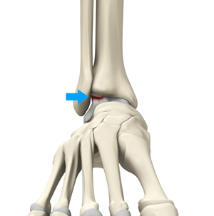
The ankle joint is formed by the articulation of the end of the tibia and fibula (shinbones) with the talus (heel bone). Osteochondral injuries, also called osteochondritis dissecans, are injuries to the talus bone. It is characterized by damage to the bone as well as the cartilage covering it. Sometimes, the lower end of the tibia or shinbone may also be affected.
What are the Causes of Osteochondral Ankle Injuries?
Osteochondral injuries are most often caused by trauma to the ankle joint, such as with ankle sprains. Some cases may not have any previous history of ankle injury and may be caused by local osteonecrosis or a metabolic defect.
What are the Symptoms of Osteochondral Ankle Injuries?
The symptoms of osteochondral ankle injuries include:
- Localized pain of the ankle joint
- Tenderness and swelling of the ankle joint
- Difficulty in weight-bearing
- Locking of the ankle
How are Osteochondral Injuries of the Ankle Diagnosed?
Osteochondral injuries are diagnosed by a physical examination, and X-ray and CT and MRI scans. Plain X-ray images can reveal other fractures, bone spurs, and narrowing of the joint. A CT scan helps identify any bony fragments and cysts, but is not very helpful to visualize bone edema or cartilage defects. MRI is the best imaging modality, which helps to visualize the cartilage and bone lesions as well as bone edema.
What are the Treatment Options for Osteochondral Ankle Injuries?
Nonsurgical or surgical treatment may be recommended for the management of osteochondral injuries of the ankle joint.
Nonsurgical treatment with immobilization, restricted weight-bearing and physical therapy may be ordered to help the bone and cartilage to heal, and improve muscle strength, mobility and coordination.
Surgical treatment is recommended for severe injuries and comprises of debridement (removal) of the damaged cartilage and removal of any loose bodies. Some of the most commonly used surgical techniques include:
- Microfracture or drilling of the lesion
- Grafting of cartilage and bone
- Fixation of the fragments with the help of screws